Cardiovascular System Mind Map
University · Physiology · Updated 31 October 2026
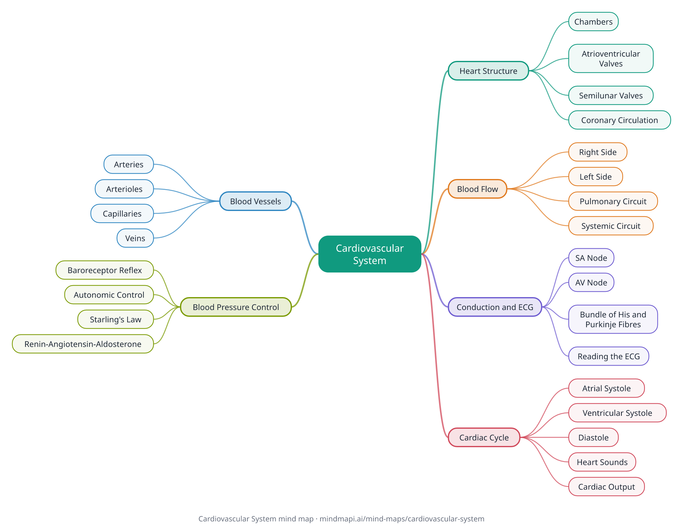
This cardiovascular system mind map puts the core physiology on one page: the structure of the heart, the route blood takes through it, the conduction system and the ECG, the cardiac cycle, the blood vessels, and how blood pressure is controlled. Every branch is explained in the notes below the map.
It is pitched at first and second-year medicine, nursing, biomedical science and allied health courses. Mind maps suit anatomy and physiology well: a 2025 systematic review found that mind maps and concept maps improved preclinical medical students' results in most of the trials it included, especially for retention.
Download this mind map
Print the map with all the notes as a PDF revision sheet, or save the map as an image. Free to use for revision and in class.
Cardiovascular System mind map explained
Heart Structure
A muscular pump with four chambers and four valves.
Chambers
Two atria receive blood; two ventricles pump it out. The left ventricle has the thickest wall because it pumps blood around the whole body.
Atrioventricular Valves
The tricuspid valve (right) and the mitral, or bicuspid, valve (left) stop backflow into the atria. Chordae tendineae and papillary muscles stop them turning inside out.
Semilunar Valves
The pulmonary and aortic valves stop blood flowing back into the ventricles.
Coronary Circulation
The coronary arteries, the first branches of the aorta, supply the heart muscle (myocardium) itself.
Blood Flow
A double circulation: the pulmonary circuit to the lungs and the systemic circuit to the body.
Right Side
Venae cavae → right atrium → tricuspid valve → right ventricle → pulmonary valve → pulmonary arteries → lungs. Carries deoxygenated blood.
Left Side
Pulmonary veins → left atrium → mitral valve → left ventricle → aortic valve → aorta → body. Carries oxygenated blood.
Pulmonary Circuit
Low pressure, so fluid is not forced into the lungs. The pulmonary arteries are the only arteries carrying deoxygenated blood.
Systemic Circuit
High pressure, to push blood through the whole body.
Conduction and ECG
The heart's own electrical system sets the rhythm (it is myogenic).
SA Node
The natural pacemaker in the right atrium. Its impulse spreads across both atria, which contract.
AV Node
Delays the impulse by about a tenth of a second, so the ventricles fill before they contract.
Bundle of His and Purkinje Fibres
Carry the impulse down the septum and through the ventricle walls, so the ventricles contract from the apex upwards.
Reading the ECG
- P wave: atrial depolarisation
- QRS complex: ventricular depolarisation (atrial repolarisation is hidden in it)
- T wave: ventricular repolarisation
- PR interval: time from atrial to ventricular depolarisation, including the AV node delay
Cardiac Cycle
One heartbeat: contraction (systole) and relaxation (diastole).
Atrial Systole
The atria contract and top up the ventricles, which are already mostly filled passively.
Ventricular Systole
The ventricles contract. The AV valves close, pressure rises with all valves shut (isovolumetric contraction), then the semilunar valves open and blood is ejected.
Diastole
The ventricles relax. The semilunar valves close, pressure falls (isovolumetric relaxation), then the AV valves open and the ventricles fill.
Heart Sounds
The first sound, "lub", is the AV valves closing. The second, "dub", is the semilunar valves closing.
Cardiac Output
cardiac output = heart rate × stroke volume. At rest, roughly 70 beats per minute × 70 mL ≈ 5 litres per minute.
Blood Vessels
Each type of vessel is built for its job.
Arteries
Thick, elastic, muscular walls to carry blood at high pressure away from the heart. Elastic recoil smooths the flow.
Arterioles
Small arteries whose smooth muscle controls resistance and directs blood flow. They are the main site of resistance.
Capillaries
Walls one cell thick, for exchange of oxygen, nutrients and waste with the tissues.
Veins
Thin walls, a wide lumen and valves. Blood returns at low pressure, helped by the skeletal muscle pump.
Blood Pressure Control
Mean arterial pressure = cardiac output × total peripheral resistance. A typical resting value is around 120/80 mmHg.
Baroreceptor Reflex
Stretch receptors in the carotid sinus and aortic arch signal the cardiovascular centre in the medulla, which adjusts the heart and vessels within seconds.
Autonomic Control
Sympathetic nerves raise heart rate and contractility and constrict vessels. The vagus nerve (parasympathetic) slows the heart.
Starling's Law
More venous return stretches the ventricle more, so it contracts more forcefully and stroke volume rises.
Renin-Angiotensin-Aldosterone
Longer-term control. The kidneys release renin; angiotensin II constricts vessels and triggers aldosterone, which makes the kidneys retain sodium and water, raising blood volume.
How to revise with this mind map
- Trace the blood. Practise following blood from the venae cavae back to the aorta, naming every chamber, valve and vessel in order, until you can do it without notes.
- Link the ECG to the cardiac cycle. For each wave, say what is happening electrically and what the chambers and valves are doing at the same time.
- Learn the equations and use them: cardiac output = heart rate × stroke volume, and mean arterial pressure = cardiac output × total peripheral resistance. Practise predicting what happens when one value changes.
- Think in reflexes. For blood pressure, follow each control loop from the receptor to the effect and back, from the fast baroreceptor reflex to the slower renin-angiotensin-aldosterone system.
- Make it yours. Build your own cardiovascular map in Mindmapi from your lecture notes, and ask the AI about any branch you are unsure of. The answer is saved into that branch's notes for next time.
Frequently asked questions
What is the order of blood flow through the heart?
Venae cavae, right atrium, tricuspid valve, right ventricle, pulmonary valve, pulmonary arteries, lungs, pulmonary veins, left atrium, mitral valve, left ventricle, aortic valve, aorta, then the body.
What do the waves on an ECG show?
The P wave is atrial depolarisation, the QRS complex is ventricular depolarisation, and the T wave is ventricular repolarisation. Atrial repolarisation happens during the QRS complex, so it is not seen separately.
How is cardiac output calculated?
Cardiac output = heart rate × stroke volume. At rest that is roughly 70 beats per minute × 70 mL, or about 5 litres per minute.
Why is the left ventricle wall thicker than the right?
The left ventricle pumps blood through the high-pressure systemic circulation to the whole body, while the right ventricle only pumps it to the nearby lungs at low pressure.
How does the body control blood pressure?
In the short term through the baroreceptor reflex, which adjusts heart rate and vessel diameter through the autonomic nervous system. In the longer term through the kidneys and the renin-angiotensin-aldosterone system, which changes blood volume.
How do I make my own cardiovascular system mind map?
Put the cardiovascular system in the centre and add branches for heart structure, blood flow, conduction, the cardiac cycle, blood vessels and blood pressure. To save time, paste your lecture notes into Mindmapi and it will build a first draft of the map for you to edit.
Turn your own notes into a mind map
Upload your class notes, a PDF or a lecture recording and Mindmapi builds the map for you. Your first map is free.
Try It Free


